Breast Fat Grafting and Syringe Liposuction
During consultations for breast fat grafting, we are often asked, "How do you take out the fat for grafting? Do you simply put back the fat that comes out during liposuction?" In fat grafting, how the fat is harvested matters just as much as how it is injected.
In this article we will explain, in order, how much fat is needed for breast and buttock fat grafting, the history and principles of syringe liposuction, the method used to harvest that fat, and why we at Venus Clinic use syringe liposuction for large-volume fat grafting.

How Much Fat Does Fat Grafting Require?
Unlike facial fat grafting, the breasts and buttocks require a much larger volume of grafted fat. That is why the harvesting method deserves separate thought.
Breasts — 200–300 mL per side
For breast fat grafting, we inject about 200–300 mL per side. This is similar to the volumes reported in the medical literature.
A study of 105 patients who had breast augmentation by fat grafting reported an average of 205 mL (120–250 mL) injected per side in one session, and noted that in thin patients who received less than 150 mL per side because there was not enough fat to harvest, the improvement was not obvious.[1] A review article gives the usual range as 100–300 mL per side.[3]
| Report | Volume injected per side (one session) | Remarks |
|---|---|---|
| 105 cases of breast augmentation by fat grafting (2014)[1] | Average 205 mL (120–250 mL) | Improvement not obvious below 150 mL |
| Study measuring volume with 3D imaging[2] | Average 205±45 mL | Volume retention of 65% at 6 months |
| Usual range in a review article[3] | 100–300 mL | Injected in small amounts across multiple layers |
Buttocks — about 1 cm in size, yet the shape changes
For the buttocks, even after grafting this much fat, the reported increase in size is about 1 cm. In a prospective study in which an average of 287 mL was grafted per side, the thickness of the subcutaneous fat measured by ultrasound increased by 0.66–0.86 cm.[4]
The number looks small, but that is because the fat is spread evenly over a wide area. Even so, the actual shape can change noticeably. This is because the areas filled by grafting and the waist and flank lines that are contoured while harvesting the fat change together.
By the Numbers
Buttocks: if 287 mL is spread evenly over an area of about 250 cm², 287 ÷ 250 ≒ 1.1 cm. Even assuming that all of the grafted fat remains, the increase in thickness is about 1 cm.[4]
Breasts: to graft 250 mL into each side, 250 × 2 = 500 mL of fat is needed. With 60 mL syringes, 500 ÷ 60 ≒ 8.3, which means nine or more syringes must be filled. Results vary by individual.
The History of Syringe Liposuction
Syringe liposuction is a method of harvesting fat using the negative pressure created by pulling back the plunger of a syringe instead of an aspirator (machine). It may sound like a new method, but it is an old technique that has developed almost side by side with the history of liposuction itself.

The 1970s — Fat begins to be suctioned by machine
The idea of removing fat by "suction" came from the Fischers, father and son, in Italy around 1975. They built a motorized suction cannula with a rotating blade behind the opening, connected it to a vacuum device, and published their results in the late 1970s.[5][6]
In 1977 the equipment became simpler: with a Karman cannula and aspirator, it became possible to perform the same operation as Fischer. In 1978 Kesselring introduced a sharp suction cannula, but instruments that cut tissue had the problem of blood and lymph fluid collecting.[5]
Later, Illouz and Fournier of France made the blunt-tipped cannula widely known.[6] Fournier also spread the method of crisscrossing tunnels from several directions to reduce surface irregularities. Cannulas at the time were 6–10 mm in diameter, much thicker than those used today.
September 1985 — An accidental discovery that began with a 5 mL syringe
Syringe liposuction was discovered by Fournier by accident. It happened in September 1985, seven years after Fischer first demonstrated machine liposuction in Paris.[5]
To hold a small 5 mL syringe firmly, he gripped the barrel and the plunger together, with the plunger pulled all the way back. When he moved the needle back and forth in that state, pieces of fat entered the syringe. Two things became clear from this: it is the back-and-forth movement that detaches the fat, and the negative pressure serves to carry the detached fat into the syringe.
When the fat obtained this way was sent for pathology examination, the answer came back that the fat tissue was not damaged. The thought that undamaged fat, harvested under sterile conditions without contact with air, could be put back where it is needed arose here. In that sense, syringe liposuction was paired with fat grafting from the very beginning.
The arrival of the 60 mL syringe and dedicated cannulas
Fournier soon began using a suction cannula attached to a 60 mL syringe. To eliminate the empty space inside the syringe, he filled it with a small amount of normal saline in advance; this made the plunger easier to pull, and the harvested fat contained less blood than fat removed by machine.
After about two years, manufacturers began making dedicated instruments. Two designs appeared — one in which the cannula passes through the tip of the syringe (French type) and one fitted onto the outside of the syringe (Tulip type) — along with a lock to hold the pulled plunger in place. Fournier wrote that "after starting to use the syringe in 1985, I never went back to the aspirator."[5]
Tumescent anesthesia, and on to fat grafting
In 1987, when Klein of the United States published the tumescent technique, bleeding in liposuction was greatly reduced.[6] With less bleeding, larger volumes could be handled, and cannulas became progressively thinner.
In the 1990s, starting from the observation that fat harvested by syringe was clear and could be grafted, "reduced negative pressure liposuction" was proposed, suggesting that the negative pressure be lowered in machine suction as well.[7]
On the fat grafting side, Coleman established a method of harvesting fat with a 10 mL syringe and injecting it with a 3 mL syringe in very small amounts in multiple directions.[8] This method, which became the basis of fat grafting today, also started its harvesting with a syringe.
| Period | What happened |
|---|---|
| Around 1975 | The Fischers, father and son, begin suctioning fat with a motorized cannula and a vacuum device |
| 1977–1978 | Simpler equipment, Fischer's demonstration in Paris, spread of the blunt cannula |
| September 1985 | Fournier accidentally discovers that fat can be harvested with a syringe |
| 1985–1987 | Use of the 60 mL syringe; French-type and Tulip-type dedicated cannulas and the plunger lock appear |
| 1987 | Klein publishes the tumescent technique |
| 1990s | Reduced negative pressure liposuction proposed; Coleman establishes syringe harvesting and small-volume dispersed grafting |
The Principles of Syringe Liposuction
The buffer effect created by saline
Machine suction takes place in a vacuum, while syringe suction takes place in liquid. The saline placed in the syringe beforehand acts as a buffer, absorbing the shock between the fat tissue and the negative pressure. Fournier named this as the key difference of the syringe and explained that it is a condition that cannot be created with a machine.[5]
Low negative pressure and fat cells
The negative pressure in a syringe is created once when the plunger is pulled, and it gradually decreases as the fat fills in. The textbook explains that lowering the negative pressure reduces bleeding, and recommends putting a little saline or air into the syringe to lower the pressure further.[5]
Conversely, there are reports that many fat cells are destroyed in fat harvested with high negative pressure.[9] Because fat for grafting needs living cells, the negative pressure at the harvesting stage is important.
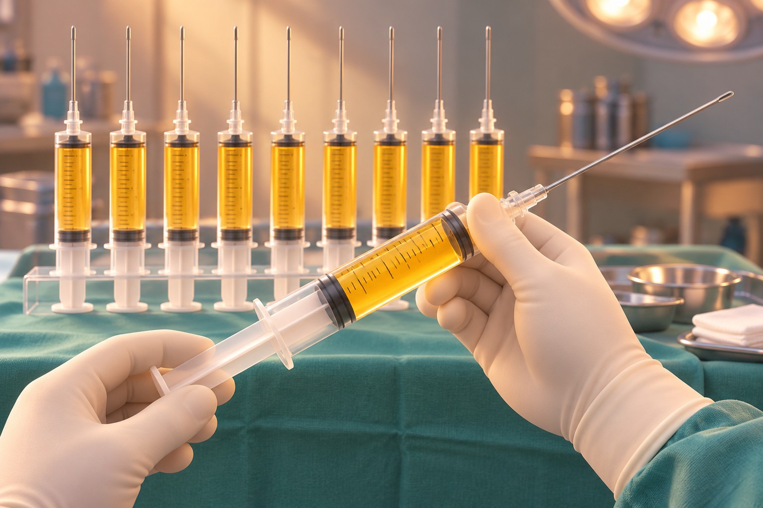
The table below summarizes the comparison Fournier presented in the textbook.[5] Please keep in mind that this is the assessment of the person who created the syringe technique.
| Item | Machine suction | Syringe suction |
|---|---|---|
| Measuring the harvested volume | Collected in a large container; hard to measure exactly | The syringe markings serve as the unit of measurement |
| Buffer effect | None | Present (pre-filled saline) |
| Damage to fat tissue | Some | Less |
| Suction speed | Same | Same |
| Noise during surgery | Yes | No |
| Grafting of the harvested fat | Described as not suitable | Can be grafted |
Comparing fat harvested by syringe with fat suctioned by SAL
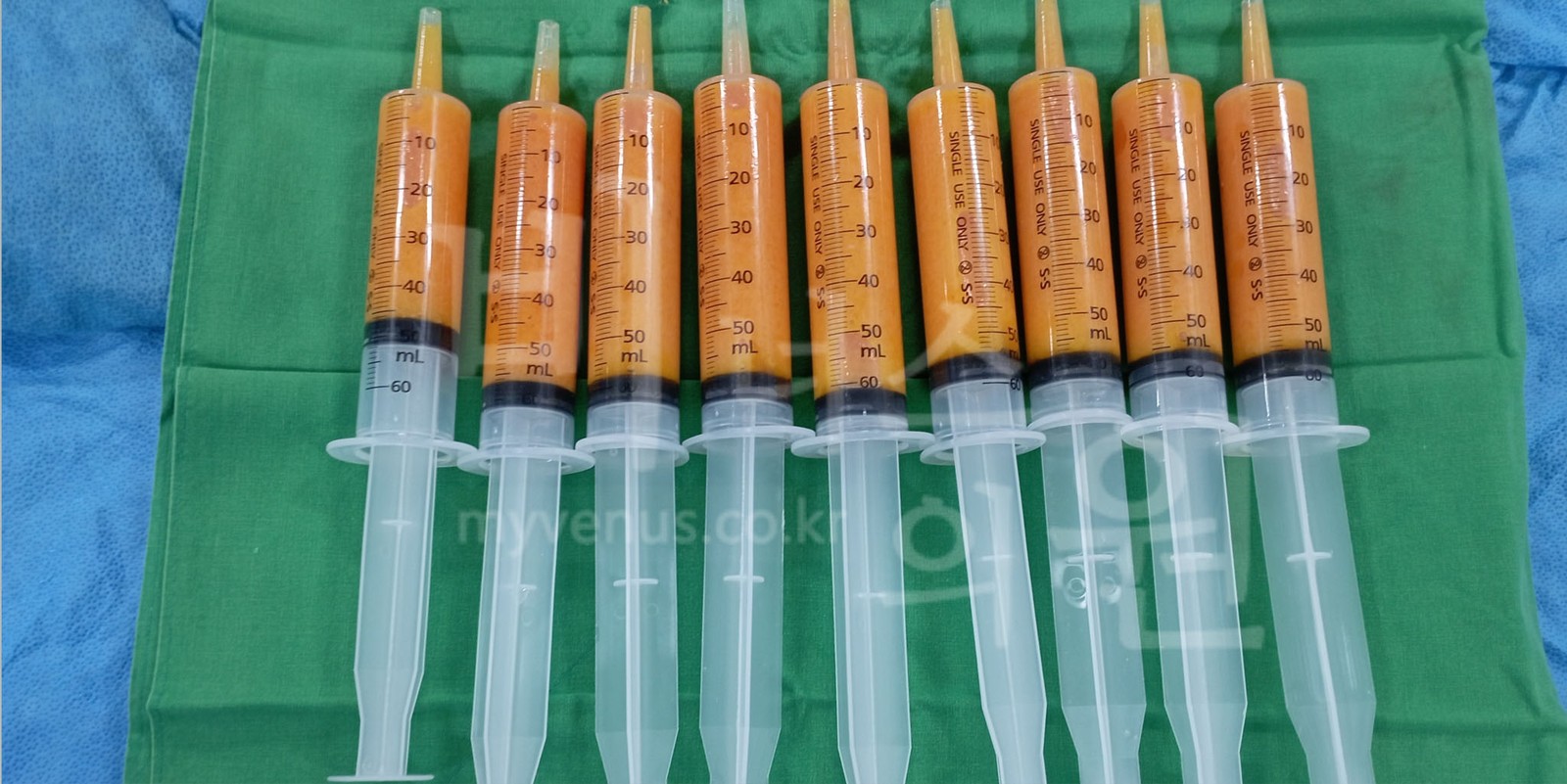
Let us compare the fat obtained by the two methods in photographs. First is fat we harvested by syringe liposuction. Each 60 mL syringe is filled with clear yellow fat.
Next is fat collected in a canister (suction bottle) by conventional suction-assisted liposuction (SAL), which uses an aspirator. The yellow upper layer is fat, and the red lower layer is tumescent fluid mixed with blood components.
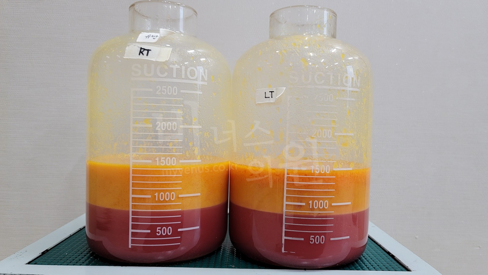
Fat harvested by syringe contains fewer blood components and has a clear yellow color, whereas with fat suctioned by machine a red fluid layer collects separately at the bottom. This is the same difference Fournier observed when he first used the syringe: "less blood is mixed in than when removed by machine." Because these photos are from different surgeries, the amounts cannot be compared directly, but the photos also suggest which is better suited as fat for grafting.
How Breast Fat Grafting Is Performed
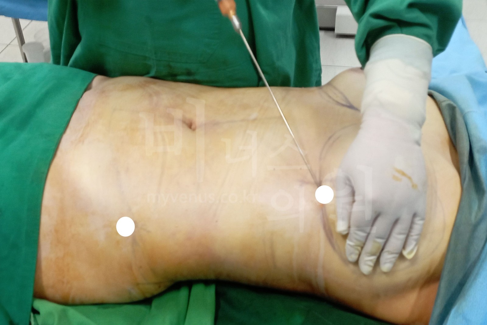
First, fat is harvested by syringe liposuction from an area with enough fat, such as the abdomen. The donor area is also contoured according to the principles of liposuction, leaving the remaining fat layer even.
After the fluid is separated from the harvested fat, the fat is grafted into the breast with a thin cannula. If a large amount is placed in one spot at once, the central part has difficulty surviving, so it is placed in small amounts across multiple layers.[1]
How Much of the Grafted Fat Remains?
In one systematic review, the volume retention of breast fat grafting was reported as 62.4% on average (44.7–82.6%).[10] However, reports differ widely depending on the measurement method, and some of the literature points to 10–50%.[2] Results vary by individual.
Limitations You Should Know
Fat grafting is not a surgery that achieves an exact desired size in a single session. Part of the grafted fat is absorbed, and in a study comparing it with implants, fat grafting required an average of two procedures.[11] Thin patients may not have enough fat to harvest in the first place.
Every surgery carries the possibility of side effects. After breast fat grafting, lumps, oil cysts, and calcification can occur, and these are checked by ultrasound examination.[12] If body weight changes significantly after surgery, the volume of the grafted fat may change as well.
Why We Use the Syringe for Large-Volume Fat Grafting
When a large volume of fat grafting is needed, such as 200–300 mL per side, we at Venus Clinic perform syringe liposuction. There are three reasons.

It helps reduce damage to the fat
With the syringe, the negative pressure is low and the saline acts as a buffer, so damage to the fat tends to be less. The more fat is grafted, the more the proportion of living fat cells matters.
The fat is used for grafting as is, without contact with air
Because the fat goes directly into the syringe, it can be harvested without contact with air, and the same syringe leads straight into preparation for grafting.
Volumes are measured by the syringe markings
The syringe markings allow the harvested volume and the volume injected on each side to be measured accurately. This gives a clear reference when placing the same amount in both breasts, or different amounts as needed.
Before-and-After Photos
These are photos of a patient whose fat was harvested by abdominal liposuction and grafted into the breasts, and all of the after photos were taken one week after surgery. Swelling and bruising are still present at this stage, so this is not the final result.
Looking at the photos, you may wonder, "Are these abdominal liposuction photos or breast fat grafting photos?" They are both. Because fat was harvested from the abdomen and moved to the breasts in a single surgery, two kinds of change are captured in one photo. That makes this a fitting case for explaining the process of harvesting fat by syringe liposuction together with breast fat grafting.
How to Read the Photos
The abdomen and flanks are where the fat was harvested — the change from liposuction.
The breasts are where that fat was placed — the change from fat grafting.
In the front and side views both areas are visible; in the back view the breasts cannot be seen, so only the waist and flanks where fat was harvested are visible.
Front view
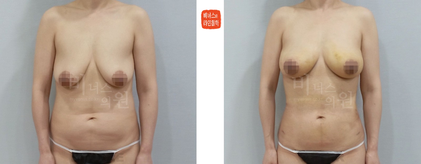
This is a case in which fat was harvested by abdominal liposuction and grafted into the breasts. The left is before surgery, and the right is the front view one week after surgery.
Changes in the abdomen — Before surgery, the lower abdomen protruded forward and the flanks bulged outward, so the waistline was close to a straight line. In the after photo, the protrusion of the lower abdomen appears reduced and the waistline appears to curve inward. Because this is where the fat was harvested, bruising and small incision marks remain.
Changes in the breasts — Before surgery, the upper part of the breasts had little volume and they appeared to sag downward. In the after photo, the upper part is filled and the overall shape appears rounder. The yellow bruising on the upper inner breast occurred during the grafting process and fades over time. At one week the size includes swelling, and part of the grafted fat will be absorbed.
| Information on the Posted Liposuction Before-and-After Photos | |||
|---|---|---|---|
| - Same person | Surgery performed at Venus Clinic; same person | - Date taken | Taken around 2025; comparison at 1 week after surgery |
| - Same conditions | Edited to the same posture and size as closely as possible | - Side effects | https://en.myvenus.co.kr/should_know/38861 |
Left side view
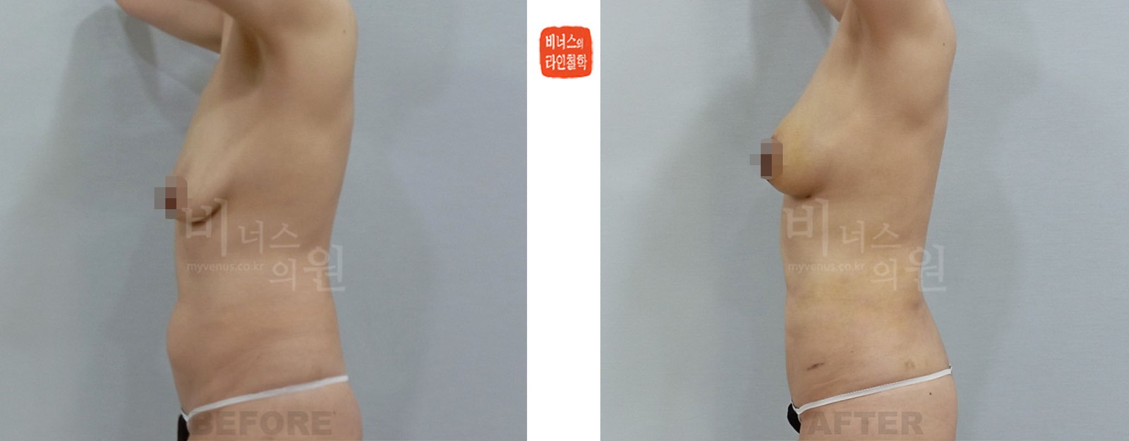
This is the left side view of a case in which fat was harvested by abdominal liposuction and grafted into the breasts.
Changes in the abdomen — Seen from the side, the line of the lower abdomen that protruded forward before surgery appears flatter in the after photo.
Changes in the breasts — The breasts, which lay flat and low before surgery, appear to project forward more in the after photo. The change from grafting is easier to see in the side view than in the front view.
| Information on the Posted Liposuction Before-and-After Photos | |||
|---|---|---|---|
| - Same person | Surgery performed at Venus Clinic; same person | - Date taken | Taken around 2025; comparison at 1 week after surgery |
| - Same conditions | Edited to the same posture and size as closely as possible | - Side effects | https://en.myvenus.co.kr/should_know/38861 |
Back view
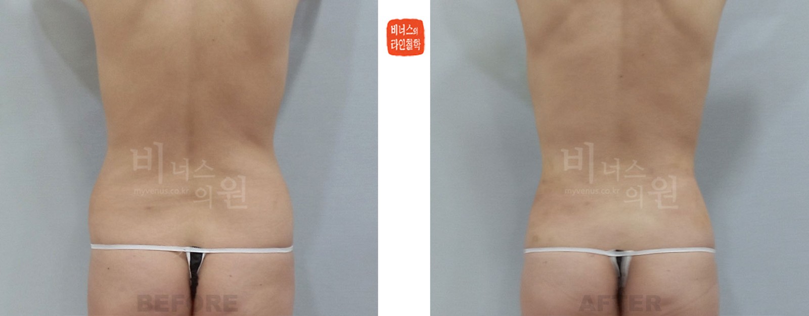
This is the back view of a case in which fat was harvested by abdominal liposuction and grafted into the breasts.
Changes in the abdomen — The flanks that protruded on both sides of the waist before surgery appear reduced in the after photo, and the line running from the back to the waist appears to curve inward. The back view shows only the area where fat was harvested.
Changes in the breasts — The breasts are not visible from the back, so this cannot be assessed in this photo.
| Information on the Posted Liposuction Before-and-After Photos | |||
|---|---|---|---|
| - Same person | Surgery performed at Venus Clinic; same person | - Date taken | Taken around 2025; comparison at 1 week after surgery |
| - Same conditions | Edited to the same posture and size as closely as possible | - Side effects | https://en.myvenus.co.kr/should_know/38861 |
Right side view
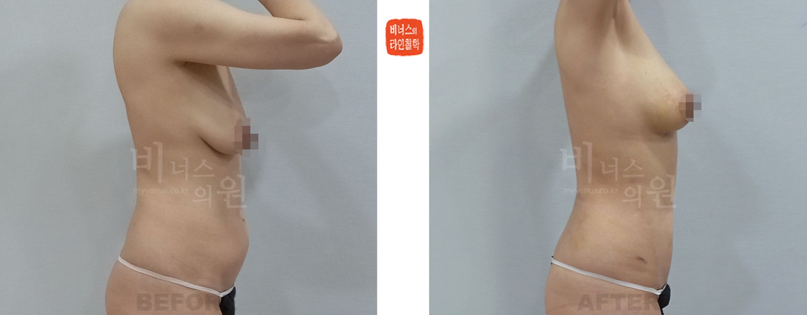
This is the right side view of a case in which fat was harvested by abdominal liposuction and grafted into the breasts.
Changes in the abdomen — The side contour in which the lower abdomen protruded forward before surgery appears gentler in the after photo.
Changes in the breasts — Before surgery, the upper part of the breast was hollow and the breast sat low; in the after photo, the upper part is filled and the line seen from the side appears rounder.
| Information on the Posted Liposuction Before-and-After Photos | |||
|---|---|---|---|
| - Same person | Surgery performed at Venus Clinic; same person | - Date taken | Taken around 2025; comparison at 1 week after surgery |
| - Same conditions | Edited to the same posture and size as closely as possible | - Side effects | https://en.myvenus.co.kr/should_know/38861 |
In Closing
What matters most in fat grafting is transferring living fat with as little damage as possible. Syringe liposuction began with one surgeon's accidental discovery in 1985 and has been refined for fat grafting from the start. At Venus Clinic, we harvest fat with this method when a large volume of fat grafting is needed, as with the breasts.
References
- Li FC, Chen B, Cheng L. Breast augmentation with autologous fat injection: a report of 105 cases. Ann Plast Surg. 2014;73 Suppl 1:S37-42.
- Wang CL, Luan SS, Panayi AC, et al. Methods used for evaluation of volume retention rate in autologous fat grafting for breast augmentation: a systematic review. Chin Med J. 2019;132(18):2223-2228.
- Aad S, Hakim R, Saad R, et al. Fatal fat embolism in cosmetic surgery: Comparing gluteal fat grafting to other lipofilling procedures. JPRAS Open. 2026;51:475-484.
- Swanson E. Prospective controlled study of buttock fat transfer using ultrasound and photographic measurements. Plast Reconstr Surg Glob Open. 2016;4(5):e697.
- Fournier PF. Reduction syringe liposculpturing. In: Shiffman MA, Di Giuseppe A, eds. Liposuction: Principles and Practice. Springer; 2006. Chapter 30 and others.
- Klein JA. Tumescent Technique: Tumescent Anesthesia & Microcannular Liposuction. Mosby; 2000. Chapter 1.
- Elam MV, Packer D, Schwab J. Reduced negative pressure liposuction (RNPL): Could less be more? Int J Aesthet Restor Surg. 1997;3(2):101-104.
- Yu NZ, Huang JZ, Zhang H, et al. A systemic review of autologous fat grafting survival rate and related severe complications. Chin Med J. 2015;128(9):1245-1251.
- Bellini E, Grieco MP, Raposio E. The science behind autologous fat grafting. Ann Med Surg (Lond). 2017;24:65-73.
- Groen JW, Negenborn VL, Twisk JW, et al. Autologous fat grafting in cosmetic breast augmentation: a systematic review on radiological safety, complications, volume retention, and patient/surgeon satisfaction. Aesthet Surg J. 2016;36(9):993-1007.
- Gentile P. Breast implants or lipofilling in augmentation mammoplasty? A randomized, open-label, controlled trial. Aesthetic Plast Surg. 2026;50(3):1044-1056.
- Shida M, Chiba A, Ohashi M, et al. Ultrasound diagnosis and treatment of breast lumps after breast augmentation with autologous fat grafting. Plast Reconstr Surg Glob Open. 2017;5(12):e1603.
Dr. Hakjun Kim, Director, Venus Clinic